Animal Cell Mitochondria Diagram - Diagram Cell Diagram Biology Full Version Hd Quality Diagram Biology Ritualdiagrams Itfpontederadevitalia It : Mitochondria have their own dna.
byCharles Marrujo-
0
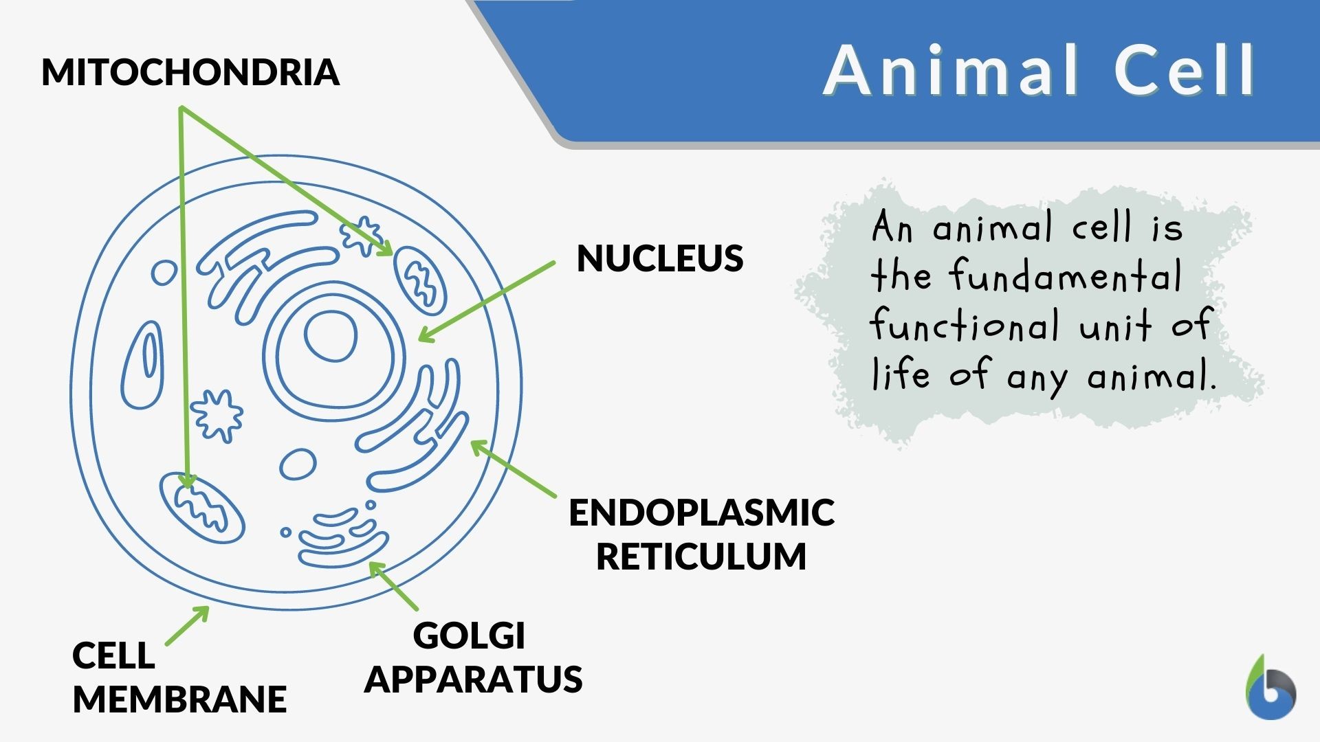
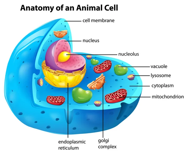
Animal Cell Mitochondria Diagram - Diagram Cell Diagram Biology Full Version Hd Quality Diagram Biology Ritualdiagrams Itfpontederadevitalia It : Mitochondria have their own dna.. The various cell organelles present in an animal cell are clearly marked in the animal cell diagram provided below. Inside the mtdna d.in the intermembrane space This occurred over a long process of millions of years, and now the mitochondria inside the cell cannot live separately from it. The proteins of bcl2 family regulates the release of cytochrome c from the inner and outer membrane of mitochondria which, once in cytoplasm, causes activation of other apoptotic signals, finally causing cell death. The cells like muscle cells, liver, indicate the higher rate of atp utilization in these areas.
See full list on studiousguy.com See full list on bbc.co.uk Animal cells have a basic structure. Its size ranges from 0.5 to 1.0 micrometre in diameter. These membranes are made of phospholipid layers, just like the cell's outer membrane.
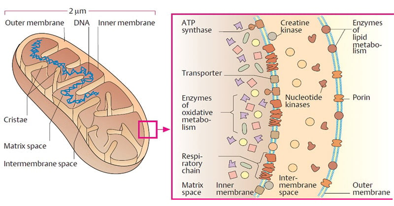
Animal Cell Definition And Examples Biology Online Dictionary from www.biologyonline.com We all keep in mind that the human physique is quite problematic and one way i discovered to comprehend it is by way of the style of human anatomy diagrams. Aug 12, 2021 · a typical animal cell will have on the order of 1000 to 2000 mitochondria. Cristae, which are the foldings of the inner mitochondrial membrane, increase the surface area of the inner membrane. Outer mitochondrial membrane(the outer layer of the mitochondrial covering) the outer membrane contains pores like structure called porins through which, small, uncharged particles can pass inside and out of the mitochondria. These membranes are made of phospholipid layers, just like the cell's outer membrane. This occurred over a long process of millions of years, and now the mitochondria inside the cell cannot live separately from it. There are many shreds of evidence to support the endosymbiotic theory of mitochondria, for example, it has its own circular dna just like prokaryotic cells. Mitochondria have their own dna.
Mitochondria divide by binary fission, independently of the cell they live in.
The cells like muscle cells, liver, indicate the higher rate of atp utilization in these areas. An animal cell is the smallest unit that makes up the varied tissues of animal species. These membranes are made of phospholipid layers, just like the cell's outer membrane. The outer membrane and the inner membrane are made of proteins and phospholipid layers. For example, mitochondria have their own dna that is separate from the dna in the cell's nucleus. This set of proteins is called an electron transport chain. Mitochondria are known as thepowerhouse of the cell because it is responsible for generating energy currency in the form of atp which is later utilized by the cell for performing various functions. Mar 23, 2021 · animal cell: Its size ranges from 0.5 to 1.0 micrometre in diameter. The citric acid cycle reduces nicotinamide adenine dinucleotide (nad+) to nadh. D.the genome is similar to that of bacterial dna. Inner mitochondrial membrane(the inner layer of mitochondrial covering) this is the site for the process of electron transport chain (etc). Electrons from nadh travel through protein complexes that are embedded in the inner membrane of the mitochondria.
Within the inner membrane b. For large, charged molecules to pass, special transmembrane proteins are required as they can not pass through the porins. Animal cells vary in different shapes and size and perform specific functions. Mar 23, 2021 · animal cell: Mitochondria also play a very essential role in the process of apoptosis in mammalian cells.
Mitochondria Definition Structure Functions And Diagram from microbenotes.com A group of cells assemble together to form tissues and eventually to organs and organ systems. This diagram shows the structure of a mitochondrion. See full list on studiousguy.com Its size ranges from 0.5 to 1.0 micrometre in diameter. The various cell organelles present in an animal cell are clearly marked in the animal cell diagram provided below. The folds increase surface area of the membrane, which is important because the inner membrane holds the proteins involved in the electron transport chain. Mitochondria divide by binary fission, independently of the cell they live in. Endosymbiotic theory states that the mitochondria and chloroplast are a result of endocytosis (engulfment) of the aerobic bacteria (prokaryote) by a eukaryotic cell.
The outer membrane covers the surface of the mitochondrion, while the inner membrane is located within and has many folds called cristae.
Mitochondria contain its own ribosome, therefore, it can synthesis its own proteins but majorly they are encoded by nucleus only (99%). Mitochondria has a vital role in uptake and release of ca2+ which maintains the concentration of calcium in the cytoplasm of the cells. Storing calcium d.all of the above 2. See full list on biologydictionary.net It is also where many other chemical reactions take place to carry out the mitochondria's many functions. The size of the mitochondria and prokaryotic cells are almost similar. Where is the mitochondrial matrix located? These membranes are made of phospholipid layers, just like the cell's outer membrane. For large, charged molecules to pass, special transmembrane proteins are required as they can not pass through the porins. Mitochondria reproduce through binary fission. More images for animal cell mitochondria diagram » Mitochondria are the power plants of the cell. The population of all the mitochondria of a given cell constitutes the chondriome.
Within the inner membrane b. The folds increase surface area of the membrane, which is important because the inner membrane holds the proteins involved in the electron transport chain. Mitochondria have two membranes, an outer membrane and an inner membrane. Where is the mitochondrial matrix located? The citric acid cycle, or krebs cycle, takes place in the mitochondria.
420 Mitochondria Vector Images Free Royalty Free Mitochondria Vectors Depositphotos from st.depositphotos.com It is the main site for atp synthesis and therefore it is called a powerhouse of the cell (oxidative phosphorylation). Aug 12, 2021 · a typical animal cell will have on the order of 1000 to 2000 mitochondria. Mitochondria reproduce through binary fission. An increased surface area creates more space for more reactions to occur, and increases the mitochondria's output. You received your mtdna from your mother, and you can only pass it on if you are a female who has a child. The size of the mitochondria and prokaryotic cells are almost similar. Cells are made up of different parts. The amount of mitochondria in a cell depends on how much energy that cell needs to produce.
It is the main site for atp synthesis and therefore it is called a powerhouse of the cell (oxidative phosphorylation).
It is also where many other chemical reactions take place to carry out the mitochondria's many functions. The citric acid cycle, or krebs cycle, takes place in the mitochondria. The multivesicular bodies (mvbs) and the exosome secretion pathway were observed in mrcs. More images for animal cell mitochondria diagram » It is easier to describe these parts by using diagrams: Storing calcium d.all of the above 2. The population of all the mitochondria of a given cell constitutes the chondriome. Mar 23, 2021 · animal cell: These membranes are made of phospholipid layers, just like the cell's outer membrane. In mature rbcs, mitochondria are absent in order to create more space for haemoglobin for oxygen binding. The outer membrane is more permeable compared to the inner membrane 1. The idea that mitochondria evolved this way is called endosymbiotic theory. Animal cells vary in different shapes and size and perform specific functions.